New MRI technique maps fluid velocity distribution in the brain
The tool could help researchers study the brain’s waste clearing system and its links to Alzheimer’s Disease or other neurodegenerative disorders.
The tool could help researchers study the brain’s waste clearing system and its links to Alzheimer’s Disease or other neurodegenerative disorders.
Experts
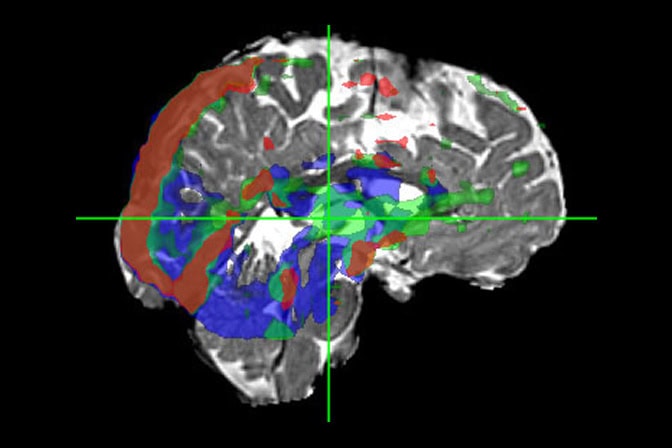
A new MRI technique called Velocity Spectrum Imaging can map fluid movement in the human brain within a 3D pixel, according to a University of Michigan Engineering study published in Magnetic Resonance in Medicine. The non-invasive approach does not need injectable or consumable contrast agents to sharpen the image.
The method has many potential applications, but the current focus is to gather a clearer image of the glymphatic system, which moves fluid through the brain, to help researchers understand its involvement in neurodegenerative disorders like Alzheimer’s Disease.
“I think this technique has the potential to be another tool to monitor and to use as a biomarker for water exchange and water movement—in the brain and beyond,” said Luis Hernandez-Garcia, a research professor of biomedical engineering at U-M and corresponding author of the study funded by the National Institutes of Health.
The glymphatic system acts as the brain’s waste management system, flushing out cellular waste with cerebrospinal and interstitial fluid. If the system is not working properly, waste proteins called amyloid-beta and tau proteins build up and disrupt cell communication. Excessive buildup of these waste proteins is a hallmark of Alzheimer’s Disease and other neurodegenerative disorders.
Current MRI techniques used to capture fluid movement, like phase contrast MRI, measure average fluid velocity for each 3D pixel, called a voxel. While this resolution works for other parts of the body, averaging misses detail in the brain where multiple velocities or directions coexist in a small space.
For example, within the same voxel of a brain MRI scan, two water channels (perivascular spaces) may cross. When averaged, the flows of water molecules in opposite directions cancel each other out. Additionally, blood flowing through capillaries coexists in the same tiny space as cerebrospinal fluid moving through the surrounding perivascular channels, frequently at different speeds in different directions.
To get a better understanding of the full spectrum of fluid movement at the voxel level, the researchers leveraged specialized magnetic pulses to label water molecules based on how fast they were moving.
A typical MRI uses a mathematical tool called a Fourier transform to determine where a signal came from in space to construct a 2D or 3D image. Here, the Fourier transform is also used to determine how fast the signal was moving to create a graph for each voxel—showing how much fluid is stationary, moving slowly or rushing past.
The researchers first confirmed that Velocity Spectrum Imaging captured accurate readings from a custom-build flow phantom—a phantom is a man-made object that has known dimensions and properties and is used to verify that the imaging system works properly. In this case, the device pumps water through tubes at a known rate and with known distribution.
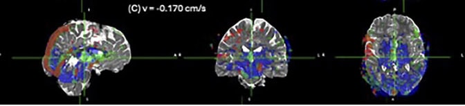
When tested on five human participants, the technique successfully mapped 3D fluid velocity distribution in the brain. Velocity bands identified anatomical landmarks like the ventricles and cerebral aqueduct, the main thoroughfares for cerebrospinal fluid.
While the study serves as a proof-of-concept for the technique, the researchers must overcome technical limitations before it reaches practical clinical use.
Improving the resolution will help Velocity Spectrum Imaging capture a wider range of fluid movement. Currently, the scanning rate is too slow to be practical in the clinic. It is also barely sensitive enough to detect ultra-low velocity of perivascular spaces of the glymphatic system.
“Right now, we think it may be useful for monitoring the functionality of the glymphatic system and help understand Alzheimer’s disease and develop new therapies. There are some technical issues that we still need to overcome to speed up the acquisition and reduce artifacts, but I’m very optimistic,” said Hernandez-Garcia.
This study was funded by the National Institutes of Health (R01NS112233, R21EB03251401A1).