Nano-wrinkles and actuation break up infectious biofilms on implants
The active antibacterial platform improved tissue response in mice and could one day help prevent infections caused by implanted medical devices in humans.

The active antibacterial platform improved tissue response in mice and could one day help prevent infections caused by implanted medical devices in humans.

Michigan Engineering is advancing
critical technologies for a healthier world
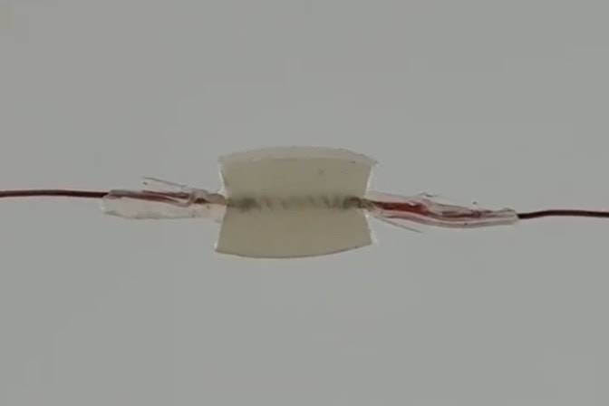
A new implantable device developed by a University of Michigan Engineering team prevented biofilm growth and improved tissue outcomes in mice. The researchers pass an electrical current through wires to heat the soft robotic coil embedded in a silicone square, causing it to heat and contract. When implanted, squeezing this silicone square pops bacteria off the nano-wrinkled surface. The study moves closer towards clinical use in humans to prevent infection-causing biofilms from forming on joint replacements, pacemakers, catheters and more. Credit: Min and Nam Laboratories, University of Michigan Engineering.
A silicone square with a coil embedded in the center has wires protruding from either side. At 0:05 and 0:10, the coil contracts and compresses the silicone square inwards, like squishing a marshmallow between two fingers. After each compression, the square slowly releases back to its original size.
A nano-wrinkled surface combined with a tiny soft actuator can break up infection-causing bacterial sheets, called biofilms, that form on medical implants, according to a University of Michigan Engineering study published in Advanced Functional Materials.
When implanted in mice, the platform—called DARTS (dynamically actuated reconfigurable topographical surface)—reduced bacterial burden and improved the surrounding tissue response.
“This work brings us a step closer to translating our basic biointerface research towards medical use in humans,” said Jouha Min, an assistant professor of chemical engineering at U-M and co-corresponding author of the study.
In humans, the platform could be adapted for medical devices like joint replacements, pacemakers and catheters, which are prone to biofilm surface growth, which can lead to hard-to-treat antibiotic-resistant infections.
“After years of studying how surface topographies shape cell-material interactions in the lab, we’re now building those principles into an implant that actively controls those interactions in the body,” said Min.
The research team built the antibacterial surface with a bottom-up nanofabrication process, stacking layers on top of one another. A pre-stretched, flexible plastic film forms the base. Next, the plastic is repeatedly dipped in alternating solutions with a positive or negative electrical charge, adhering layers as opposite charges attract.
The coated plastic is placed in an oven at 130 C, causing the pre-stretched plastic base to shrink back to its original size, much like a Shrinky Dink. The number of coatings drives the size of the wrinkles. A single coating in each solution creates nano-wrinkles, with wavelengths shorter than 400 nanometers. Dipping the plastic 20 times creates micro-wrinkles with about 2 micron (2,000 nanometers) wavelengths.
The sharp waves of the nano-wrinkles broke apart both gram-negative and gram-positive bacteria, suppressing biofilm coverage. The micro-wrinkles failed to suppress bacterial growth as bacteria could settle in between the larger grooves.
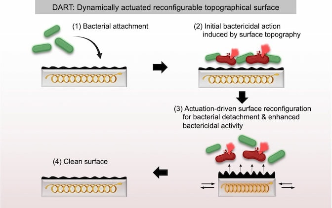
To further enhance the bacterial suppression, the researchers integrated a soft robotic actuator into the nano-wrinkled surface. The actuator consists of a tiny metallic spring embedded in a small, soft silicone block with wires extending outwards. The actuator sits at the bottom of the device and the nano-wrinkled surface on top with a thin silicone cushion in the middle.
Passing an electrical current through the spring causes it to heat and contract, squeezing the top nano-wrinkles together and popping bacteria off the surface. The insulation surrounding the spring and protruding wires keeps the heat inside the device to prevent harm when implanted.
When cultured with bacteria, the dynamic actuation combined with nano-wrinkles produced near-complete disruption and removal of mature biofilms. The system combatted biofilm buildup over repeated challenges and maintained its shape through 200 actuation cycles.
“What makes this approach particularly exciting is that we are moving beyond passive antibacterial surfaces toward a system that can actively respond to bacterial buildup. By integrating mechanical actuation with engineered surface topography, we can physically disrupt established biofilms and potentially prevent persistent infections on medical implants,” said Sungmin Nam, an assistant professor of mechanical engineering at U-M and co-corresponding author of the study.
To test the platform in living animals, the team implanted the DARTS devices bearing bacteria (Staphylococcus aureus) beneath the skin in mice. After seven days, the researchers collected the device and a sample of surrounding tissue.
The DARTS platform was actuated 20 times immediately after surgery and again on days one, three and five. The actuated DARTS platform had markedly lower bacterial contamination and better local tissue outcomes compared to all controls.
The team also showed that a near-infrared laser can actuate the device wirelessly. This could make the approach more practical for clinical use, where an external light wand might one day clear bacterial buildup without surgery.
“Rather than relying on passive coatings, our surface physically removes bacteria when needed. That opens a new path toward preventing implant infections,” said Min.
This research was supported by the National Institutes of Health (R35GM157070; 1R03AG093279-01A1), National Science Foundation (2441718), V Foundation V Scholar Award (V2023-002), Rogel Cancer Center Discovery Award, and the Korean-American Scientists and Engineers Association Young Investigator Grant.
The device was studied at the Michigan Center for Materials Characterization and animal research was conducted with the support of the Unit for Laboratory Animal Medicine, both of which are operated and maintained with support from indirect cost allocations in federal grants.