Next generation neural probe leads to expanded understanding of the brain
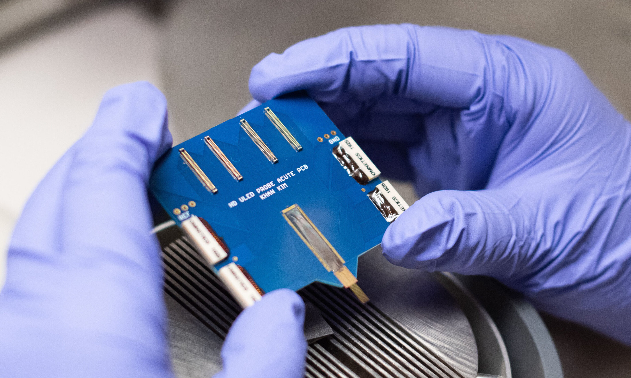
The hectoSTAR probe, with 128 stimulating micro-LEDs and 256 recording electrodes integrated in the same neural probe, was designed for some stellar brain mapping projects
The hectoSTAR probe, with 128 stimulating micro-LEDs and 256 recording electrodes integrated in the same neural probe, was designed for some stellar brain mapping projects

A newly-developed neural probe with an unprecedented number of micro-LEDs and recording sites integrated on the same neural device is enabling neuroscientists to gain new knowledge into how the brain operates. The 128 μLEDs and 256 recording electrodes on the hectoSTAR probe allow neuroscientists to track interactions across different regions of the brain.
“With the hectoSTAR probe, we could address some questions that we were not able to answer before,” said Mihály Vöröslakos, neuroscientist at New York University and first author of a new study. “Having technology that no one else has gives a rare opportunity to be the first to test these devices.”
Having more channels allows neuroscientists to not only record a greater area of the brain, but also to record the interactions between different areas, and this may lead to important insights into treating neurological disorders.
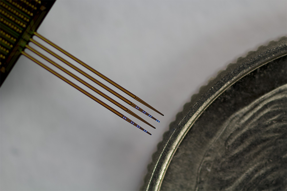
The hectoSTAR probe can cover a region spanning the surface of a rodent’s brain, called the cortex, all the way down to the hippocampus in the deep brain. Comprised of four 30-μm thick silicon micro-needle shanks, it is capable of recording from about 1 mm2 area (1.3 mm x 0.9 mm), up to as deep as 6 mm inside the brain.
After two years of development by researchers at the University of Michigan, it worked the first time in real-life conditions.
The hectoSTAR probe was placed in the CA3 region of the hippocampus of a live mouse. Light was passed through one or more LEDs, and the neuronal response was monitored by the recording electrodes. The team discovered that the light elicited a response not only in neighboring cells in the same CA3 region, but that it also impacted the neurons in the neighboring CA1 region. The CA3 and CA1 regions are 600 microns apart, or a little more than half a millimeter.
“With this tool, we can perturb the neural system in a very precise spatial and temporal manner,” said Vöröslakos. “The hectoSTAR device, and in general the micro LED probe, is an amazing tool to enable these experiments.”
Advancing scientific research takes patience – and often a high level of teamwork and cooperation. Already familiar with earlier versions of the neural probe developed by Prof. Euisik Yoon’s team at the University of Michigan, Vöröslakos spent two years in Yoon’s lab at Michigan as a Kavli scholar to ensure the new technology could address very specific scientific questions.
In 2015, Yoon’s team reported the first-of-a-kind neural probe that can record activities from multiple neurons and stimulate the neurons’ activities at a nearly single-cellular resolution. Those probes contained 12 μLEDs and 32 recording electrodes. In 2020, Yoon’s team, led by former doctoral student Kanghwan Kim (MSE PHD EE ‘15, ‘20), introduced a more practical microLED probe that allows high-quality neural signal recording during LED stimulation by removing the large amount of noise that LED stimulation had generated in the recorded neural signal.
“That work paved the way to scaling it up,” said Yoon. “There were a number of engineering challenges to overcome, but the result has enabled neuroscientists to increase their understanding of the global connectivity of the brain. We are excited about this new chip.”
The new μLEDs, measuring 8 x 11 micrometers, are about half the area of the previous generation. Kanghwan Kim, a lead researcher of the hectoSTAR team, put theory into practice to get sufficient light output to stimulate the neurons.
“Nothing was trivial in terms of shrinking things down,” said Kim.
Finally, increasing the number of optical stimulation sites to 128 was a bit of a nightmare, said Yoon. To deal with the control issue, his team designed an open-source multi-channel μLED controller system to individually control the light being sent to each μLED, and then to process the incoming information from the recording electrodes.
“We can precisely control which LEDs get illuminated, as well as the intensity of the light, which has the ability to stimulate only a few neurons close to the LED, and then record how the signal is propagating to other parts of the brain,” said Yoon.
Increasing the number of LEDs and electrodes by an order of magnitude took time. “After spending so much time in the cleanroom, I did have confidence that it would work,” smiled Kim. “It was exciting seeing those micro LEDs come to life.”
“Without this probe, we would need to put multiple probes into the brain to get the same information. Even then, we were limited by how much weight the animal could carry,” explained Vöröslakos.
Based on the results of the hectoSTAR electrode, Vöröslakos is running followup experiments, and is already asking for the next generations of the devices.
In response, Yoon has set his current team of researchers to achieve two important enhancements to the hectoSTAR: flexibility of the probes, and a greatly miniaturized headstage board. These improvements would allow the rodent to move freely with longevity while its brain was being simultaneously stimulated and recorded.
Researchers from other universities have already been visiting Yoon’s lab to learn how to implant his neural probes and collect the data.
Vöröslakos is a member of The Neuroscience Institute at New York University. Antonio Fernandez-Ruiz, corresponding author of the study, is an Assistant Professor at Cornell University and a former postdoctoral researcher of György Buzsáki. Prof. Buzsáki, corresponding author of the study, is the Biggs Professor of Neuroscience at NYU and decades-long collaborator with Yoon’s group. Yoon and Wise, corresponding author and co-author, are co-founders of NeuroLight Technologies, a for-profit manufacturer of neurotechnology.
The hectoSTAR was developed in the Lurie Nanofabrication Facility. Research was supported by the National Institutes of Health, National Science Foundation, a Brain & Behavior Research Foundation Young Investigator Award, a Fulbright Scholarship, and the Kavli Foundation.
Study: HectoSTAR μLED Optoelectrodes for Large-Scale, High-Precision In Vivo Opto-Electrophysiology, Mihály Vöröslakos, Kanghwan Kim, Nathan Slager, Eunah Ko, Sungjin Oh, Saman S. Parizi, Blake Hendrix, John P. Seymour, Kensall D. Wise, György Buzsáki, Antonio Fernández-Ruiz, and Euisik Yoon, Advanced Science. https://doi.org/10.1002/advs.202105414