X-pinch plasma achieves radial proton acceleration for crisp imaging
Plasma pinches: From pursuits of nuclear fusion to an attractive point source of accelerated protons for proton radiography.

Plasma pinches: From pursuits of nuclear fusion to an attractive point source of accelerated protons for proton radiography.

Advancing nuclear energy
from discovery to deployment
Protons accelerated in a radial direction were discovered and used for the first time from pinch plasmas—current-carrying plasma columns compressed by their own magnetic field—according to a study led by the Czech Technical University in Prague and University of Michigan Engineering. This research was primarily funded by the Grant Agency of the Czech Republic and the U.S. Department of Energy.
The researchers accelerated protons to 3 mega electron volts (MeV) on relatively small-scale devices operating at a 400 kiloamperes (kA) peak current. This expands access to proton radiography, a technique for imaging the ultra-fast evolution of electric and magnetic fields in plasma, once limited to sophisticated, expensive and often massive laser facilities like the OMEGA and OMEGA-EP laser systems at the University of Rochester’s Laboratory for Laser Energetics.
“This new proton radiography capability will lead to a better understanding of pinch physics in general, potentially leading to advances in nuclear fusion research,” said Ryan McBride, a professor of nuclear engineering and radiological sciences and electrical and computer engineering at U-M and a senior author of the study published in Physical Review Letters and Physics of Plasmas.
The results show that a broader range of laboratories can now conduct proton radiography experiments, opening new opportunities in high-energy-density plasma research and fusion science.
The first plasma pinch, called a Z-pinch, was investigated in the search for controlled nuclear fusion. In this setup, a cylindrical column of ionized gas (plasma) is aligned with the z-axis of a cylindrical coordinate system. A pulsed-power generator then sends a massive burst of electrical current through the plasma column. The direction of the current is parallel to the z-axis, hence the name “Z-pinch.”
The intense current generates a strong magnetic field that surrounds the plasma. This field interacts with the current, crushing the plasma inwards, towards the z-axis (the axis of symmetry). This compression usually results in a massive burst of X-rays, emitted in all directions, and a burst of accelerated protons emitted in the axial z-direction—the same direction as the current. In recent times, Z-pinches have been adapted for imaging applications as well as studying nuclear fusion and astrophysical phenomena.
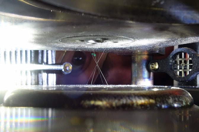
For imaging applications, Z-pinches can produce blurry images if the source of radiation is spread out into a long plasma column. In this study, however, the research team used a hybrid X-pinch configuration, which emits protons from a short pinch column to produce a clearer image. In an X-pinch, the current and plasma compression is focused to the small crossing point of an X-shaped structure. This can generate sub-millimeter, or even micron-sized radiation sources.
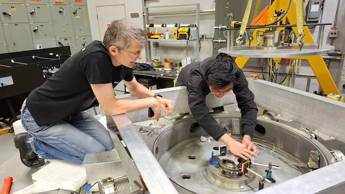
Experiments were conducted on two pulse generators, the MAIZE facility at U-M and the XP facility at Cornell University, both of which operated at a maximum of 400 kA. To create the hybrid X-pinch plasma, the research team strung a polyethylene (CH2) fiber, just 30 micrometers thick (about half as thick as a human hair), between two solid conical electrodes that faced one another, forming the X-shape.
As in a standard Z-pinch, the electric current pulse vaporizes and ionizes the fiber into a plasma column. The X-shape of the conical electrodes, however, focuses the magnetic pressure and forces the pinch to occur at a single, precise location.
The ultra-thin fiber disintegrates so fast that it creates an abrupt current disruption because the plasma column breaks apart and can no longer carry the current. The disruption of the electrical energy that was flowing through the plasma results in a massive buildup of voltage, higher than the machine’s initial output pulse. This accelerating voltage causes the protons to slingshot out of the plasma in a radial direction at near-relativistic speed.
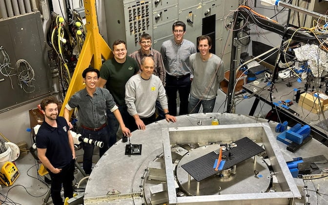
“For more than 40 years, X-pinches have been studied worldwide primarily as point-like sources of X-rays and, to a lesser extent, fast electrons and fusion neutrons,” said Daniel Klír, a professor of applied physics at the Czech Technical University in Prague and lead author of the study.
“Their potential as compact sources of accelerated ions has been largely overlooked. The key was driving the current through an extremely thin, hydrogen-rich fiber. This breakthrough was made possible through a very fruitful collaboration, combining the pulsed-power expertise of our colleagues at the University of Michigan and Cornell University with our expertise in ion acceleration and diagnostics.”
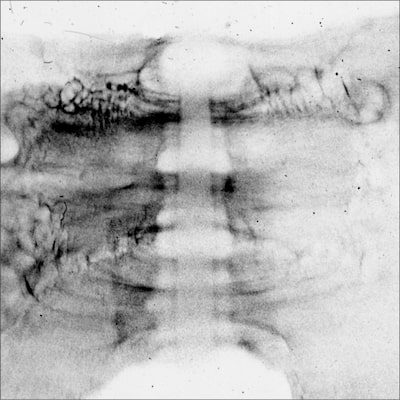
The research team demonstrated the use of radial acceleration by imaging an exploding wire placed adjacent to the X-pinch. The X-pinch was placed in the center of the machine, while a specialized ion detector was placed behind the exploding wire to capture the shadow-like proton image.
After the electrical pulse fired, the radially accelerated protons from the X-pinch captured the wire’s explosion with extreme clarity. The resulting image shows the dense center of the exploding wire, an expanding cloud of plasma, and even the turbulent structures surrounding the explosion.
Researchers at Cornell University also contributed to this study.
The University of Michigan Engineering and Cornell teams were supported by the United States Department of Energy (DE-SC0020239; DE-NA0004148; DE-NA0004027). The Czech team was supported by the Grant Agency of the Czech Republic (Grant No. 23-04679S).